
Click here to see all images
March, 2022
Case of the Month
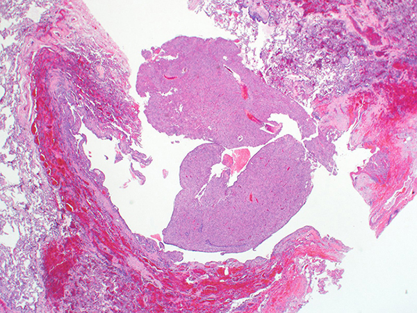
Clinical History: A 50-year-old woman with minimal remote history of smoking was found to have a 1.5 cm mildly PET avid nodule in the right lower lobe without lymphadenopathy. She underwent right lower lobe wedge resection. H&E photomicrographs of the nodule are shown in Figures 1-4 and an immunohistochemical stain for HMB-45 in Figure 5. The neoplastic cells were negative for cytokeratin AE1/AE3, S100, CD68, and SMA.
Q1. This tumor is thought to have what line of differentiation?
- Histiocytic
- Hepatocytic
- Perivascular epithlioid cell
- Renal
Q2. The histopathologic features of this tumor include all, except:
- Neuroendocrine differentiation
- Nested or sheet-like growth pattern
- Uniform round cells with clear or granular eosinophilic cytoplasm
- Subendothelial growth around thin-walled vessels
Q3. Most of these tumors harbor TFE3 gene rearrangements
- True
- False
Answers to Quiz
Q1. C
Q2. A
Q3. B
Q2. A
Q3. B
Diagnosis
PEComa of the lung
Discussion
Perivascular epithelioid cell tumors (PEComas) of the lung, formerly known as sugar tumor or clear cell tumor, are rare benign mesenchymal neoplasms with perivascular epithelioid-cell differentiation. They are part of the PEComa family along with lymphangioleiomyomatosis (LAM), angiomyolipoma, and diffuse PEComatosis. PEComas are composed of cells that are often associated with blood vessel walls and usually express melanocytic and smooth muscle markers.
Lung PEComas are often peripherally located solitary nodules and are incidentally found on chest imaging. Unlike extrathoracic PEComas, they tend to be more frequent in males. There is a wide age range, with a peak in older adults >40 years old. While most PEComas are sporadic; a small subset is associated with tuberous sclerosis. Approximately 80% of PEComas show TSC2 mutations and result in abnormal signaling through the mTOR pathway, while a minority harbor TFE3 gene fusion.
Grossly, lung PEComas are well circumscribed, with a firm, glistening red-tan cut surface, typically measuring less than 5 cm. Histologically, this tumor has a sharp border with the alveolated parenchyma and is composed of uniform round/ovoid epithelioid cells with abundant clear or granular eosinophilic cytoplasm and distinct cell borders. The tumor cells grow in sheets or organoid nests (Figures 1-4). Thin-walled sinusoidal vessels are easily identified, with tumor cells showing a subendothelial growth pattern, supporting the perivascular nature of this neoplasm (Figure 3). Malignant PEComa can be encountered in the setting of metastasis from extrapulmonary sites and is characterized by mitotic activity, marked atypia, pleomorphism, and necrosis.
Immunohistochemical stains are helpful in confirming the diagnosis and excluding other entities such as carcinoma (especially renal cell or hepatocellular), melanoma, alveolar soft part sarcoma, pure smooth muscle tumors, granular cell tumor, and myoepithelial tumors. PEComas are negative for keratins and typically express melanocytic markers, such as HMB45 (Figure 5), melan-A, and MITF, and muscle markers, such as SMA, desmin, and caldesmon. Some tumors, such as the current case, lack expression of muscle markers and ~10% are focally positive for S100.
Virtually, all PEComas of the lung are benign and cured by excision.
Take home message for trainees: Not every tumor with melanocytic differentiation is a melanoma. The lack of cytologic atypia and presence of clear or granular cytoplasm should be clues to the diagnosis.
References
1. Malinowska I, Kwiatkowski DJ, Weiss S, et al. Perivascular epithelioid cell tumors (PEComas) harboring TFE3 gene rearrangements lack the TSC2 alterations characteristic of conventional PEComas: further evidence for a biological distinction. Am J Surg Pathol 2012;36:783-4.
2. Maloney N, Giannikou K, Lefferts J, et al. Expanding the histomorphologic spectrum of TFE3-rearranged perivascular epithelioid cell tumors. Hum Pathol 2018;82:125-30.
3. Jain D, Doyle LA. PEComa of the lung. In: WHO Classification of Tumours. Thoracic tumours. Lyon (France): International Agency for Research on Cancer; 2021. pp. 172–3. (WHO classification of tumours series, 5th ed.; vol. 5). https://publications.iarc.fr/595.
Contributors
Mitra Mehrad, MD
Associate Professor
Department of Pathology, Microbiology and Immunology
Vanderbilt University School of Medicine
Nashville, TN, USA
Associate Professor
Department of Pathology, Microbiology and Immunology
Vanderbilt University School of Medicine
Nashville, TN, USA