
Click here to see all images
November, 2022
Case of the Month
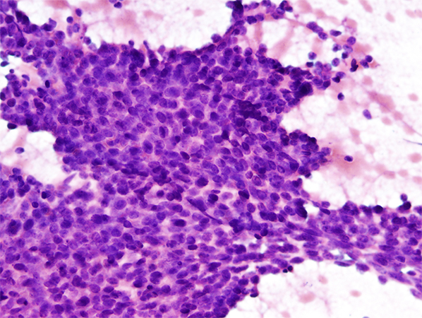
Clinical History: A 59-year old woman with a history of breast cancer presented with coughing and shortness of breath and was found to have a 1.2 cm endobronchial mass at the left bronchus. The patient underwent fiberoptic bronchoscopy and a sessile, polypoid mass was seen at the left mainstem bronchus. An endobronchial fine needle aspiration (FNA) was performed and representative images from the direct smears and cell block preparation are shown in Figures 1-5.
Q1. Which of the following histopathologic features would not be considered common for this entity?
- Basaloid cells with angulated nuclei
- Basophilic matrix-like material
- Brisk mitosis and tumor necrosis
- Cribriform architectural pattern
Q2. Which of the following would not be considered common for these tumors?
- Distant metastasis
- Infiltrative growth pattern
- Local recurrence
- Perineural invasion
Q3. What molecular alteration would be considered diagnostic for this entity?
- CRTC1::MAML2 gene fusion
- EWS::FLI1 gene fusion
- MYB::NFIB gene fusion
- NUTM1::BRD3 gene fusion
Answers to Quiz
Q1. C
Q2. A
Q3. C
Q2. A
Q3. C
Diagnosis
Adenoid cystic carcinoma
Discussion
The cytologic smears are moderately cellular and demonstrate a monotonous population of basaloid cells with oval/angulated hyperchromatic nuclei, with evenly dispersed chromatin, indistinct nucleoli and scant cytoplasm. In some areas, matrix-like material was present associated with the tumor cells, seen more prominently on the Giemsa-stained smear, and as a pale-staining material on the Pap-stained smear. Cell block sections highlighted nests of tumor cells with punched-out spaces containing basophilic matrix. Immunohistochemical stains were positive for CK7, S100, and CD117. The final diagnosis was a pulmonary adenoid cystic carcinoma.
Adenoid cystic carcinoma is a rare salivary gland-type malignant neoplasm often arising in the mainstem bronchus and presenting as an endobronchial mass lesion. The lesions are frequently detected on a computed-tomography (CT) scan and may be diagnosed using fine needle aspiration (FNA) under bronchoscopic guidance. Histologically the tumor is composed of small, angulated, basaloid cells with frequent perineural invasion. The most common pattern seen is the cribriform, showing nests of tumor cells with punched-out spaces associated with basophilic matrix. Other architectural patterns seen less commonly are tubular and solid. The differential diagnoses include any basaloid tumor, including basaloid squamous cell carcinoma and carcinoid tumors. Immunohistochemical staining can be performed to differentiate between the entities to make the diagnosis of adenoid cystic carcinoma. In addition, these tumors demonstrate MYB::NFIB gene fusions that can be helpful to establish a definitive diagnosis, especially in cases where the characteristic cribriform morphologic pattern is not present. These tumors have an infiltrative growth pattern with perineural invasion, making complete surgical resection difficult. Local recurrence is common; however, distant metastases are relatively uncommon.
Take home message for trainees: Adenoid cystic carcinoma is a rare salivary gland–type carcinoma often presenting as an endobronchial mass. It is a biphasic tumor demonstrating both ductal and myoepithelial phenotypes with small basaloid cells with angulated nuclei and associated with basophilic matrix. The MYB::NFIB gene fusion is characteristic for these tumors.
References
Doxtader EE, Shah AA, Zhang Y, et al. Primary salivary gland-type tumors of the tracheobronchial tree diagnosed by transbronchial fine needle aspiration: Clinical and cytomorphologic features with histopathologic correlation. Diagn Cytopathol. 2019 Nov;47(11):1168-1176. doi: 10.1002/dc.24285.
Monaco SE, Khalbuss WE, Ustinova E, et al. The cytomorphologic spectrum of salivary gland type tumors in the lung and mediastinum: a report of 16 patients. Diagn Cytopathol. 2012 Dec;40(12):1062-70. doi: 10.1002/dc.21733.
Molina JR, Aubry MC, Lewis JE, et al. Primary salivary gland-type lung cancer: spectrum of clinical presentation, histopathologic and prognostic factors. Cancer. 2007 Nov 15;110(10):2253-9. doi: 10.1002/cncr.23048.
Contributors
Sinchita Roy-Chowdhuri, MD PhD
Associate Professor
Department of Pathology
The University of Texas MD Anderson Cancer Center
Houston, TX, USA
Associate Professor
Department of Pathology
The University of Texas MD Anderson Cancer Center
Houston, TX, USA