
Click here to see all images
March, 2024
Case of the Month
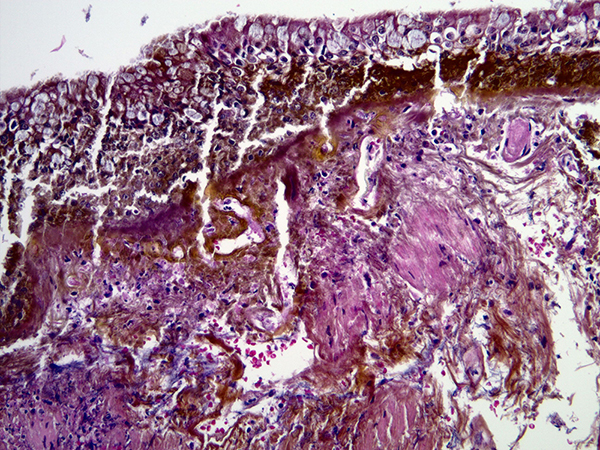
Clinical History: A woman in her 70s with a history of COPD and anemia presented to the emergency room with shortness of breath. She was diagnosed with a COPD exacerbation, and subsequently developed acute-on-chronic respiratory failure. Bronchoscopy showed copious mucoid tan-brown and thick secretions in the right middle lobe and right lower lobe. The bronchial mucosa showed yellow discoloration (Figure 1). Bronchial brushings and washings were obtained, and an endobronchial biopsy was performed. Images from the endobronchial biopsy are shown in Figure 2 (H&E), Figure 3 (H&E) and Figure 4 (Iron [Perls’] stain).
Q1. What should you (the pathologist) look for in the history in this case?
- Whether the patient is immunocompetent or immunocompromised.
- History of connective tissue disease.
- History of prior malignancy
- History of iron pill (FeSO4) intake and predisposing factors for aspiration.
Q2. This patient was taking iron (FeSO4) pills for anemia. What effect do iron pills have on the bronchial mucosa if aspirated?
- None. They are harmless.
- They cause neoplastic transformation.
- As in the GI tract, they can cause erosion and ulceration.
- They have no effect on bronchial mucosa but they do damage the alveoli.
Q3. Which of the following is true regarding iron pill aspiration.
- You should correlate clinically, and if there is no history of aspiration, you should not make this diagnosis.
- These fragments cannot be seen in cytology samples.
- In most patients with iron pill aspiration, there is no history of aspiration, and the diagnosis is rarely suspected prior to biopsy.
- Iron particles have no effect on the bronchial epithelium.
Answers to Quiz
Q2. C
Q3. C
Diagnosis
Discussion
Clinically, patients with iron pill aspiration present with cough, dyspnea, wheezing, hemoptysis, chest pain or other non-specific symptoms. Only a few individuals recall choking on or aspirating a pill. Patients invariably have a history of taking oral iron (FeSO4) pills, as in this case. Most do not recall an aspiration episode, and this diagnosis is seldom suspected clinically prior to biopsy. A minority of patients have well-defined predisposing conditions that render them susceptible to aspiration.
On imaging, the findings are non-specific; they tend to be more common in the right lung, as expected. Lobar collapse/atelectasis, patchy opacification, mucus plugging and other non-specific findings are common. Very rarely, a small endobronchial lesion with a focus of hyperattenuation (possibly representing pill fragments) may be noted within an airway on chest CT. The diagnosis is almost never suspected on imaging.
On bronchoscopy, an important clue to the diagnosis is the presence of striking yellow or brown mucosal discoloration (Figure 1), sometimes with plaque-like yellow mucosal lesions. Mucous plugging is common. In some cases, the changes are interpreted as a mass lesion. Mucosal ulceration and bronchial stenosis are noted in some cases.
The most critical factor in making this diagnosis is an endobronchial biopsy, in which iron pill particles can be easily seen within bronchial mucosa (Figures 2 and 3), provided that the pathologist is aware of the morphology of iron pill particles on H&E. These particles can also be seen in bronchial brushing samples by cytopathologists. Iron pill (FeSO4) particles are elongated, fibrillary or crystalline, yellow to brown particles that are much larger in size than hemosiderin particles. They also differ in shape. Although both iron pill particles and hemosiderin stain strongly with a Perls’ stain (Figure 4), iron pill particles are usually extracellular whereas hemosiderin is typically found within macrophages. The appearance of iron particles in bronchial mucosal biopsies is identical to their appearance in biopsies from the gastrointestinal tract. The types of damage in the bronchial mucosa are also similar to the gastrointestinal tract, mucosal erosion and ulceration being common. Some patients develop bronchial stenosis in the long run.
There is no well-defined treatment. Interventions that have been tried include bronchoscopic removal of pill fragments (only possible in relatively early cases), debridement with a Nd:YAG laser, bronchial dilatation, corticosteroids, topical mitomycin, cryotherapy, and even lung resection. The prognosis is variable, but the clinical course can be protracted in some individuals.Take home message for trainees: Learn to recognize what iron pill particles look like on endobronchial biopsies. In most cases, this diagnosis is made by pathologists, and is clinically unsuspected.
References
Henriques V, Santos DC, Lérias G, Monteiro LC. Iron pill aspiration: Cytologic and histologic findings of a potential life-threatening airway injury. A Case report and literature review. Diagn Cytopathol 2018;46:532-539.
Melillo G, Scala G, Chiummariello A, Palumbo U. Sténose de la bronche principale gauche, secondaire à l'inhalation d'un médicament à base de sulfate ferreux [Stenosis of the main left bronchus after inhalation of a drug with ferrous sulfate base]. Bronches 1975;25:184-188. French.
Xing J, Yadav R, Ntiamoah P, et al. Airway injury caused by aspiration of iron sulfate pills: a series of 11 cases. Mod Pathol 2023; 36:100347.Contributors
Department of Pathology
Cleveland Clinic
Cleveland, OH, USA